We use several different techniques in our studies that allow us to examine what and how we learn new things about language. Read below to find out more about these techniques and how we use them!
Infant looking time techniques:
Headturn Preference Procedure
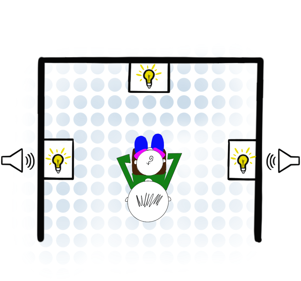
In this procedure, the infant sits on a caregiver’s lap or in a highchair, and sees a light/computer screen directly in front and at 90 degree angles to the right and left. When the infant looks to one of the side screens/lights, we play an auditory or audio-visual stimulus for as long as the infant is interested. When the infant gets bored, we turn that stimulus off, and wait for the infant to look to the other side. Once the baby looks, we start playing another stimulus. We create stimuli that we think will be more or less interesting to infants, and then see if that prediction matches how long (on average) babies are interested in listening/watching the different stimulus types.
Central Fixation & Eye Tracking
Like in the head-turn preference procedure described above, the baby sits on their caregivers’ lap/in a highchair. In this procedure, however, there is only one visual display, which is directly in front of the infant. We measure how long the infant is interested in watching/listening to different kinds of stimuli that are presented on the screen, or when they shift attention between items on a screen. We can use this procedure to see if infants discriminate between different kinds of sounds, prefer certain sound combinations over others, or if they have learned associations between sounds and objects that were implicitly presented during a learning phase.
Neuroimaging techniques:
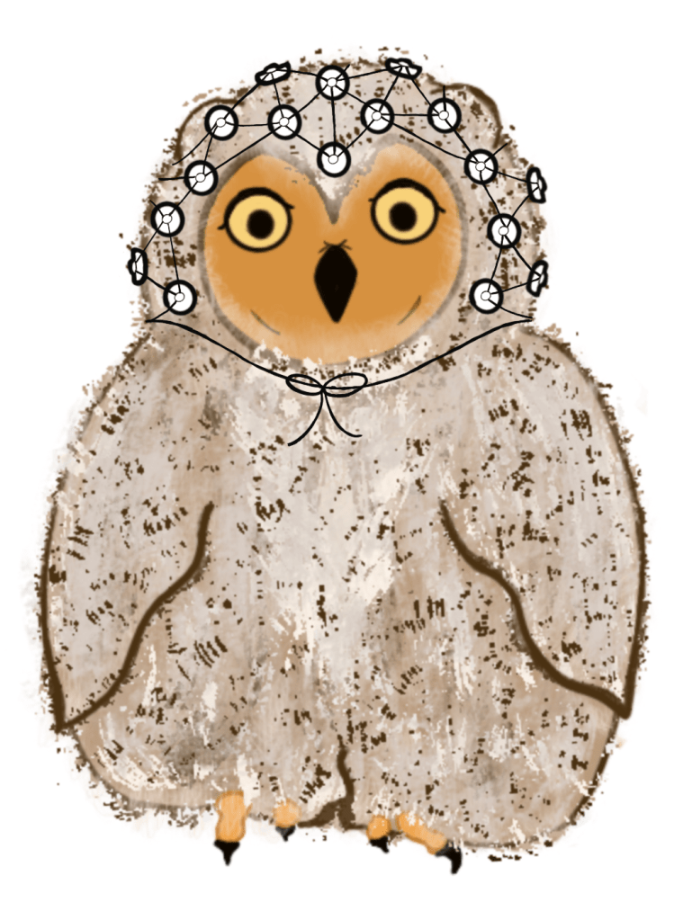
EEG
Electroencephalography (EEG) is a neuroimaging technique used to measure electrical activity within the brain. A swimcap-like hat with sewn-in electrodes that have soaked in a saline and soap solution is placed on the participant’s head and records brain activity while the participant is presented with a stimulus and/or completes a task. The solution on the electrodes allows us to better measure the brain activity, but is neither harmful nor painful. Through this technique, we can see how quickly different areas of the brain react when adults or babies are listening to language stimuli.
fNIRS
Functional near infrared spectroscopy (fNIRS) measures brain activity by tracking where blood flows through the brain. This is very similar to functional magnetic resonance imaging (fMRI), but unlike MRI, fNIRS uses near-infrared light to detect where higher levels of oxygenated blood are in the brain. When a particular area of the brain is being used, blood flow in that region becomes more oxygenated than it is at rest. To use fNIRS, we put a soft and flexible cap on the participant’s head. This cap contains a set of sensors that shine light or detect how that light is reflected back. Changes in the amount of light reflected back allow us to see which areas of the brain are being used in language processing.

